Optimum respiratory health is not possible without clearing secretions in the airway. Normally, a healthy person can get rid of these secretions in two ways: (1) mucociliary clearance system and (2) coughing. However, presence of diseases such as asthma, cystic fibrosis, cerebral palsy, dystrophy of the muscles and immunodeficiency, can cause poor lung health and inability to clear out excretions.
Inability to clear the lungs of these discharges can lead to complications and causes an individual to experience difficulty of breathing. When breathing becomes a hard work it can lead to inflammatory episodes, respiratory infections, increase mucus production and worst airway obstruction. This is where chest physiotherapy comes in. It helps in minimizing the risks of ineffective airway clearance due to obstruction.
Description
Chest physiotherapy, also known as chest physical therapy, is a group of techniques that mobilizes or loosens thick secretions in the lungs and respiratory tract. It includes postural drainage, chest percussion and vibration, coughing and deep breathing exercises.
Purpose
Performing chest physical therapy techniques expand the lungs, strengthen the muscles used for breathing, thereby improving lung function and helping one breath better. It helps treat patients diagnosed with cystic fibrosis and COPD (chronic obstructive pulmonary disease). Post-operatively, it helps in keeping lungs clear from thick secretions preventing pneumonia. Performing the techniques to a bedridden patient is critically important as helps prevent or treat atelectasis and pneumonia.
Contraindications
- Active pulmonary bleeding with hemoptysis and the immediate posthemorrhagic stage
- Fractured ribs or an unstable chest wall
- Lung contusions
- Pulmonary tuberculosis
- Untreated pneumothorax
- Acute asthma or bronchospasm
- Lung abscess or tumor
- Bony metastasis
- Head injury
- Recent myocardial infarction
- Vomiting
- Immediately after eating
Equipment Needed
- Stethoscope
- Pillows
- Tilt or postural drainage table or adjustable hospital bed
- Emesis basin
- Facial tissues
- Suction equipment
- Equipment for oral care
- Trash bag (optional)
- Sterile specimen container
- Mechanical ventilator
- Supplemental oxygen
Preparation of Equipment
- Gather equipment at the patient’s bedside.
- Set up suction equipment, if needed, and test its function.
Implementation
- Confirm patient identity.
- Explain the procedure to the patient, provide privacy and wash hands.
- Auscultate patient’s lungs to determine baseline respiratory status.
- Position patient as ordered. In generalized diseases, drainage start at the lower lobes moving on to the middle lobes and ends with the upper lobes. For localized diseases, drainage begin with the affected lobes then to the other lobes to prevent spreading of diseases to uninvolved areas.
- Instruct patient to remain in each position for 3-15 minutes. (see various position for postural drainage below) During this time, perform percussion and vibration as ordered.
- After postural drainage, vibration or percussion, instruct the patient to cough. Doing this removes loosened discharges. Instruct patient to inhale deeply though his nose and then exhale in three short buffs. Then have him/her inhale deeply again and cough through a slightly opened mouth. Three consecutive coughs are highly effective. An effective cough sounds deep, low and hollow whereas an ineffective one is high-pitched.
- Provide oral hygiene since oral secretions may have a foul taste or smell.
- Auscultate the patient’s lungs to evaluate effectiveness of therapy.
Chest Physiotherapy Techniques
- Postural Drainage
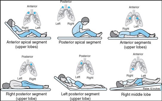
It is the sequential repositioning of the patient to encourage emptying of peripheral pulmonary discharges by gravity to the major bronchi or trachea when performed with percussion and vibration. The following are the various postural drainage position and the areas of the lungs affected:

Positions for Postural Drainage |
|
| Adult | |
| Bilateral | High fowler’s position |
| Apical segments | Sitting on side of bed |
| Right upper lobe – anterior segment | Supine with head elevated |
| Left upper lobe – anterior segment | Supine with head elevated |
| Right upper lobe – posterior segment | Side lying with right side of chest elevated on pillows |
| Left upper lobe – posterior segment | Side lying with left side of chest elevated on pillows |
| Right middle lobe – anterior segment | ¾ supine position with dependent lung in Trendelenburg position |
| Right middle lobe – posterior segment | Prone with thorax and abdomen elevated |
| Both lower lobes – anterior segment | Supine in Trendelenburg |
| Left lower lobe – lateral segment | Right side-lying in Trendelenburg position |
| Right lower lobe – lateral segment | Left side-lying in Trendelenburg position |
| Right lower lobe – posterior segment | Prone with right side of chest elevated in Trendelenburg position |
| Both lower lobes – posterior segment | Prone in Trendelenburg position |
| Child | |
| Bilateral – apical segments | Sitting on nurse’s lap leaning slightly forward flexed over pillow |
| Bilateral – middle anterior segments | Sitting on nurse’s lap, leaning against the nurse |
| Bilateral lobes – anterior segments | Lying supine on nurse’s lap, back supported with pillow |
Chart Source: Fundamentals of Nursing 3rd Edition, Potter and Perry
- Percussion
Percussion loosens and mobilizes retained secretions with cupped hands. To facilitate dislodgment of thick, tenacious secretion from the bronchial walls, the nurse claps the specific lobe or segment with cupped hands.
- Vibration
Vibration is an alternative method to percussion. Since the latter is quite uncomfortable and tiring, vibration is done to patients who are in pain, frail or recovering from thoracic surgery or trauma.
There are variety of techniques employed by nurses to treat lung diseases, included in the choices are postural drainage, suctioning and breathing exercises. The choice of therapy is based on the patient’s diagnosis and overall condition.


![Caring for Patients with Tracheostomy and Nursing Diagnoses [ Updates] tracheostomynursingprocedure](https://rnspeak.com/wp-content/uploads/2020/10/tracheostomynursingprocedure_725820712-238x178.jpg)


