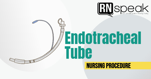
Endotracheal intubation is a very common procedure especially in the critical care unit for patients with airway problems. Patients who require mechanical ventilation needs to be intubated: either with an endotracheal tube (usually for short-term use) or a tracheostomy (long-term use).
Thus, this article would like to expand the knowledge of nurses with the common management for patients who are intubated. This article aims to provide information about:
- Indications for endotracheal intubation
- Nursing roles during the endotracheal intubation and the equipment nurses should prepare before the intubation
- How to insert oropharyngeal airway or bite block
- Basic nursing interventions for patients with an endotracheal tube
- How to perform proper endotracheal suctioning
Before we dig into the basic interventions, allow me to walk you through to the criteria behind the intubation.
Why some patients need endotracheal intubation?
Indications for Endotracheal Intubation
- Respiratory failure
- Apnea / respiratory arrest
- Inadequate ventilation(acute vs chronic)
- Inadequate oxygenation
- Chronic respiratory insufficiency with FTT
- COPD
- Airway obstruction
- Hypoventilation
- Severe hypoxemia
- Cardiac insufficiency
- Eliminate the work of breathing
- To reduce the oxygen consumption
- Neurologic dysfunction
- Impaired cognitive impairment
- Central hypoventilation and frequent apnea
- Comatose patient with GCS < 8
- Inability to protect the airway
- ABG Results
- If the patient is under the following conditions:
- Multiple trauma
- Shock
- Multi-organ failure
- Drug overdose
- Thoracic or abdominal surgery
- A long period of surgery
- Neuromuscular disorders
- Inhalation injury
So what are the things you need to prepare when the doctor says “we need to intubate the patient”? Basically, as a nurse, you should be well acquainted with the basic intubation equipment and pre medications by heart. This will come in handy when an emergency situation arises.
Your nursing checklist of intubation equipment:
- Endotracheal tubes: size 6,6.5, 7, 7.5, 8
- Laryngoscope with blades of different sizes
- Curved blades (e.g. Macintosh blades)
- Straight blades (e.g. Miller or Wisconsin blades)
- Bulbs and batteries for the laryngoscope
- Syringe 10 cc for the introduction of air/pressure to the cuff
- KY jelly or lubricating gel
- Bag valve mask
- Face mask
- Oropharyngeal airways
- Leukoplast or tape to keep ET in place (some institutions uses ET ties)
- Bite block/tongue guard
- ET holder
- Suction tip and connection tubes
- Suction machine: portable or wall-pressured
- Oxygen source
- Standby mechanical ventilator machine
- Cardiac monitor with a pulse oximeter
- Stethoscope
- IV kit
Medications and kit to prepare
- Syringe 1 cc, 3 cc, 5cc, and 10 cc
- Diazepam
- Midazolam
- Atropine
- Lidocaine
- Fentanyl
- Salbutamol for nebulization
- In-line nebulization kit
Endotracheal Intubation Procedure
Nursing roles during insertion of the endotracheal tube
It is the physician’s responsibility to insert an endotracheal tube but it doesn’t mean that nurses do not have a big role during this emergency procedure.
So what are your nurse’s roles, in the event, that this emergency happens?
- If the patient is in respiratory distress, oxygenate the patient using a bag valve mask. Attach the patient to a pulse oximeter for monitoring. Make sure to ask for reinforcement of nurses to help you in this procedure. Delegate tasks immediately (E.g. medication nurse, nurse who will assist the physician and prepare the laryngoscope, nurse who will assess the condition of the patient and checks vital signs, and etc.). One nurse cannot perform all the tasks simultaneously written below.
- Ensure that the emergency cart is accessible to the room or the area of the patient.
- If the patient has no intravenous access, immediately insert a line (or ask another nurse or intravenous therapist) for premedication purposes.
- Position the patient and the height of the bed comfortable to the physician who will insert the tube. Align the patient’s head in a neutral position. Hyperextended the head to a comfortable degree.
- Consider premedication, optional for most patients-usually given 2-3 minutes prior to induction. Prepare and administer the sedative medication as ordered by the physician.
- Prepare the laryngoscope and blades. Ensure that the batteries and bulbs are working. Ask the physician what size or type of blade he/she preferred to use.
- Assist the physician during insertion. When the tube is already in place, inflate the cuff to the desired cuff pressure using a syringe. Check the tube position and the level in the lip line (e.g. 20 cm, 21 cm, 22 cm, and 23 cm)
- Fix the tube in place partially using tape or tie, to ensure that the tube is steady. Assessment should be done first if the tube is in the correct place.
- Continue to oxygenate the patient using bag-valve or manual resuscitator.
- Verify the tube position immediately. Auscultate both lung fields. Assess if both chests are rising equally.
- Check also the pulse oximeter to assess a patient’s oxygenation.
- If the endotracheal tube is correctly placed, secure tube in position using either a leukoplast, an ET holder, or ET ties. Suction patient’s secretions as needed.
- Attach the patient to a mechanical ventilator. Check the physician’s orders for the mechanical ventilator settings.
- The physician would request a standard chest x-ray to confirm ET placement. Correspondingly, the physician would order an ABG test one hour after attaching the patient to the mechanical ventilator.
- When ABG results are out, the physician would typically adjust the mechanical ventilator settings according to the patient’s response.
Reminder: Observe the 5 moments of hand hygiene and ensure that the health care team is in their proper and complete PPEs adequate.
After the patient is intubated, what’s next? Getting the tube isn’t the end of the story, as a nurse, keeping and taking care of the patient with an endotracheal tube is another discussion. Let’s take a look at the things you can do as a nurse.
Nursing Management for patients with endotracheal tube
-
- Ensure that the required oxygen support indicated for the patient is provided.
- Assess the client’s respiratory status at least every 2 hours or frequently as indicated. Note the lung sounds and presence of secretions.
- Ensure that adequate humidity is provided to avoid feeling of dryness in the oropharynx.
- Suction secretions orally to prevent aspiration. This also decreases the risk for infection.
- Assess nasal and oral mucosa for redness and irritation.
- Secure the endotracheal tube with tape or ET holder to prevent movement or deviation of the tube in the trachea.
- Place the patient in a side-lying position or semi fowler’s if not contraindicated to avoid aspiration. Reposition patient every 2 hours. This will allow the lungs to expand better and prevent secretions stagnation.
- Ensure the ET for placement. Note lip line marking and compare it with the desired placement (18cm, 20cm, and 22cm).
- Closely monitor cuff pressure, maintaining a pressure of 20 to 25 mmHg to minimize the risk of tracheal necrosis.
- Move the oral endotracheal tube to the opposite of the mouth every 8 hours or depending on the protocol of the hospital. This is to prevent irritation to the oral mucosa.
- Provide oral care at least every 4 hours using an antibacterial or antiseptic solution. Use a bite block to avoid patient from biting down. Frequent oral care in intubated patients will decrease the risk of ventilator-acquired pneumonia.
- Use a bite block to avoid patient from biting down.
- Turn patient’s head to the side to reduce the risk for aspiration.
- Communicate frequently with the client. Give patient means to communicate using a whiteboard or communication board.
- Nutritional Consideration (Smeltzer, S., et. al, 2010)
-
- Oral feeding is contraindicated. Enteral feeding is the route for nutritional support. The most common route for enteral feeding is through a nasogastric tube.
- Do steps in preventing aspiration during feeding. Check the placing of the nasogastric tube. Place patient in high Fowler’s position.
- Ensure patient’s comfort during suctioning and other procedure that involves manipulating the endotracheal tube.
-
Nursing Care Plan for Patients with Endotracheal Tube (based on NANDA)
Nursing Diagnosis: Ineffective Airway Clearance
May be related to:
- Retained secretions; secretions in the bronchi; exudate in the alveoli; excessive mucus; airway spasm; foreign body in the airway; the presence of artificial airway
- Infection
Possibly evidenced by:
- Subjective
- Dyspnea
- Objective
- Diminished/adventitious breath sounds [rales, crackles, rhonchi, wheezes]
- Cough ineffective/absent; excessive sputum
- Changes in respiratory rate/rhythm
- Difficulty vocalizing
- Wide-eyed; restlessness
- Cyanosis
- Agitated
Desired outcomes/evaluation criteria—patient will:
- Maintain airway patency.
- Expectorate/clear secretions readily.
- Demonstrate absence/reduction of congestion with breath sounds clear, respirations noiseless, improved oxygen exchange (e.g., absence of cyanosis, ABG/pulse oximetry results within client norms).
- Verbalize understanding of cause(s) and therapeutic management regimen.
- Demonstrate behaviors to improve or maintain clear airway.
- Identify potential complications and how to initiate appropriate preventive or corrective actions.
| Nursing Intervention | Rationale |
| Assessment Assess airway patency. | Obstruction may be caused by accumulation of secretions, bronchospasm, mucous plugs and other problems. |
| Note for excessive coughing, high pressure alarms in the ventilator, increased dyspnea. | Intubated patient has ineffective cough reflex altering ability to cough. |
| Assess patient’s vital signs especially respiratory rate and rhythm. Monitor oxygen saturation using pulse oximeter. | This will serve as a baseline date. |
| Independent
Suction secretions as needed, limiting duration to less than 15 seconds. |
Suction should not be routine and duration should be limited to reduce hazard of hypoxia. |
| Reposition patient every 2 hours or more. | Promotes drainage of secretions. |
| Collaborative
Administer bronchodilators (e.g. Sabutamol 1 neb 250 mcg/neb) through in-line nebulization. Perform chest physiotherapy after the administration. Suction patient after nebulization. |
Promotes ventilation and removal of secretions. |
Nursing Diagnosis: Ineffective Breathing Pattern
May be related to:
- Anxiety; [panic attacks]
- Pain
- Perception/cognitive impairment
- Fatigue; [deconditioning]; respiratory muscle fatigue
- Hyperventilation; hypoventilation syndrome [alteration of client’s normal O2:CO2 ratio (e.g., lung diseases, pulmonary hypertension, airway obstruction, O2 therapy in COPD)]
Possibly evidenced by:
- Subjective
- Difficulty of breathing
- Objective
- Dyspnea; orthopnea
- Bradypnea; tachypnea
- Alterations in depth of breathing
- Timing ratio; prolonged expiration phases; pursed-lip breathing
- Decreased minute ventilation/vital capacity
- Decreased inspiratory/expiratory pressure
- Use of accessory muscles to breathe; assumption of three-point position
- Altered chest excursion; [paradoxical breathing patterns]
- Nasal flaring; [grunting]
- Increased anterior-posterior diameter
Desired outcomes/evaluation criteria—patient will:
- Establish a normal/effective respiratory pattern as evidenced by absence of cyanosis and other signs/symptoms of hypoxia, with ABGs within client’s normal/acceptable range.
- Verbalize awareness of causative factors.
- Initiate needed lifestyle changes.
- Demonstrate appropriate coping behaviors.
| Nursing Intervention | Rationale |
| Independent
Asess respiratory rate and depth by listening to lung sounds. |
Respiratory rate and rhythm changes are early warning signs of impending respiratory difficulties. |
| Note muscles used for breathing(sterno-cleidomastoid, diaphragmatic) and retractions/flaring of nostrils | These signify an increase in work of breathing |
| Position client with proper body alignment(semi-fowler’s position) | This is for good lung excursion and chest expansion |
| Ensure that oxygen delivery system is applied to the patient, the appropriate amount of oxygen is delivered | This provides adequate oxygenation to prevent patient from desaturation
|
| Pace and schedule activities providing adequate rest periods | This prevents dyspnea resulting from fatigue
|
| Encourage sustained deep breaths by emphasizing slow inhalation, holding end inspiration) | These promote deep inspiration |
| Teach client appropriate deep breathing and coughing techniques | These facilitate adequate clearance of secretions
|
| Collaborative
Administer oxygen at lowest concentration indicated |
For management of underlying pulmonary condition and respiratory distress. |
Nursing Diagnosis: Pain, Acute
May be related to:
- Physical agents, e.g. suctioning, manipulated endotracheal tube
- Psychological manifestations, e.g. anxiety, fear
Possibly evidenced by:
- Subjective
- Coded report since patient is unable to talk
- Changes in appetite
- Objective
- Observed evidence of pain
- Guarding behavior; protective gestures; positioning to avoid pain
- Facial mask; sleep disturbance (eyes lack luster, beaten look, fixed or scattered movement, grimace)
- Expressive behavior (e.g., restlessness, moaning, crying, vigilance, irritability, sighing)
- Distraction behavior (e.g., pacing, seeking out other people and/or activities, repetitive activities)
- Change in muscle tone (may span from listless [flaccid] to rigid)
- Diaphoresis; change in blood pressure/heart rate/respiratory rate; pupillary dilation
- Self-focusing; narrowed focus (altered time perception, impaired thought process, reduced interaction with people and environment)
Desired outcomes/evaluation criteria—patient will:
- Report pain is relieved/ controlled.
- Follow prescribed pharmacological regimen.
- Verbalize non-pharmacologic methods that provide relief.
- Demonstrate use of relaxation skills and diversional activities, as indicated, for individual situation
| Nursing Intervention | Rationale |
| Pain Management
Independent Investigate reports of pain. Note changes in degree (use scale of 0–10) and site. |
Helpful in assessing need for intervention; may indicate developing complications. |
| Monitor vital signs, note nonverbal cues, e.g. muscle tension, restlessness. | May be useful in evaluating verbal comments and effectiveness of interventions. |
| Provide quiet environment and reduce stressful stimuli, e.g. noise, lighting, constant interruptions. | Promotes rest and enhances coping abilities. |
| Place in position of comfort and support joints, extremities with pillows/padding. | May decrease associated bone/joint discomfort. |
| Reposition periodically and provide/assist with gentle ROM exercises. | Improves tissue circulation and joint mobility. |
| Provide comfort measures (e.g. massage, cool packs) and psychological support (e.g. encouragement, presence). | Minimizes need for/enhances effects of medication. |
| Review/promote patient’s own comfort interventions, e.g. position, physical activity/nonactivity, and so forth. | Successful management of pain requires patient involvement. Use of effective techniques provides positive reinforcement, promotes sense of control, and prepares patient for interventions to be used after discharge. |
| Evaluate and support patient’s coping mechanisms. | Using own learned perceptions/behaviors to manage pain can help patient cope more effectively. |
| Encourage use of stress management techniques, e.g. relaxation/deep-breathing exercises, guided imagery, visualization; Therapeutic Touch. | Facilitates relaxation, augments pharmacological therapy, and enhances coping abilities. |
| Assist with/provide diversional activities, relaxation techniques. | Helps with pain management by redirecting attention. |
| Collaborative:
Administer medications as indicated: Analgesics, e.g. acetaminophen (Tylenol) |
Given for mild pain not relieved by comfort measures. Note: Avoid aspirin-containing products because they may potentiate hemorrhage. |
| Antianxiety agents, e.g., diazepam (Valium), lorazepam (Ativan). | May be given to enhance the action of analgesics/opioids. |
So what is oropharyngeal airway or bite block for?
This is used when you need to suction secretions from the patient’s airway. Also, it keeps the air passageways open when they are obstructed by the tongue or secretions.
- Prepare a size of oropharyngeal airway appropriate to the patient’s size and age.
- Quickly place the client in a Semi-Fowler’s position or supine position if possible.
- Immediately put on clean gloves and face mask. Follow the standard precautions at all times.
- Hold the lubricated airway by the outer flange, with the distal end pointing up.
- Open the client’s mouth and insert the airway along the top of the tongues.
- When the distal end of the airway reaches the soft palate at the back of the mouth, rotate the airway 180 degrees downward, and slip it past the uvula into the oral pharynx. The oropharynx may be suctioned as needed by inserting the suction catheter alongside the airway.
- Remove after the oropharyngeal airway after use.
Patients with endotracheal tubes do not have the ability to cough-out their secretions or clear their airway. So it is our responsibility as nurses, to maintain a patent airway to the patient. One basic thing that we should learn is suctioning.
Your nursing checklist of how to perform endotracheal suctioning
Equipment
- Suction catheter and suction connecting tube
- Normal Saline Irrigation
- Suctioning machine or device: wall or portable
- Oxygen source
- Personal protective equipment
- Pulse oximeter
- Stethoscope
- Bag valve or manual resuscitator
Procedure
- Check the guidelines or standard procedure of your unit for suctioning patient with endotracheal tube.
- Prepare all needed equipment. Position all supplies so that they are easily accessible. Check the suction setup for correct functioning.
- Explain the procedure to the client. Explain why you need to suction the secretions and how it could help the patient breathe easier.
- Assess patient first. Auscultate patient’s lung fields for abnormal breath sounds (e.g. crackles, wheezing, and stridor).
- Attach patient to continuous pulse oximeter monitoring device.
- Observe stand precaution at all times. Wear personal protective equipment. Perform hand washing.
- Attach suction connection tube to the suction tip. Use the appropriate suction tip size.
- Ensure that wall or portable suction is turned on (no higher than 120 mmHg). Set a vacuum setting according to the policy of your unit.
- Hyper-oxygenate patient to 100% with the manual resuscitator for 2 – 5 minutes.
- Introduce a catheter until a restriction is met or until you can stimulate the cough reflex. You can also suction patient while he/she is coughing.
- Withdraw the catheter slowly while applying intermittent suction. Suction should not be applied for more than 15 seconds.
- Upon completion of suctioning, withdraw catheter, ensuring that tip is completely withdrawn from airway.
- Repeat the suctioning process until the patient’s airway is clear.
- Use suction tip is for single-use. Discard after use.
- Discard personal protective equipment and wash hands.
- Evaluate patient’s condition by auscultating the lung fields and by monitoring patient’s oxygenation using a pulse oximeter.
Conclusion
As a nurse, it comes in handy if you are well aware of the basic interventions or management during an emergency, most especially when it concerns airway management. Time is always of the essence.
However, though this article may provide a basic background, it is always a nurse’s duty to be acquainted with the hospital’s protocols or guidelines for standard procedures.
Moreover, aside from being knowledgeable, every nurse should always apply what they have learned. Besides, nursing is not merely textbooks. Nursing is an applied science.
References:
- Ashton, R. & Burkle, C. 2004. Endotracheal Intubation by Direct Laryngoscopy. American Thoracic Society. Retrieved at http://www.thoracic.org/professionals/clinical-resources/critical-care/critical-care-procedures/endotracheal-intubation-by-direct-laryngoscopy.php last May 4, 2015
- Michael D., et. al. 2002. Guidelines for Emergency Tracheal Intubation Immediately Following Traumatic Injury. Eastern Association for the Surgery of Trauma. East Practice Management Guidelines Workgroup. St. Elizabeth
- Doenges, M., Moorhouse, M. & Murr, A. 2006. Nursing Care Plans Guidelines for Individualizing Client Care Across the Life Span. F.A. Davis Company, Philadelphia. 7th edition.
- Health Center 1044 Belmont Ave. Youngstown, OH retrieved at https://www.east.org/Content/documents/…/intubation.pdf last May 4, 2015
- Kozier, B. et. al. 2008. Kozier and Erb’s Fundamental of Nursing Concepts, Process, and Practice. Pearson Education Inc. Prentice Hall. Upper Saddle River, New Jersey. 8th edition.
Myers, E. (2006). RNotes: Nurse’s Clinical Pocket Guide. F. A. Davis Company. Philadelphia. 2nd edition. - Silvestri, L. (2008). Comprehensive Review for the NCLEX-RN Examination. Saunders Elsevier. 4th edition.
- Smeltzer, S., Bare, B., Hinkle, J., Cheever, K. (2010). Brunner & Suddarth’s Textbook of Medical-Surgical Nursing. Lippincott Williams & Wilkins. 12th edition
- Other sources: http://www.surgeryencyclopedia.com/Ce-Fi/Endotracheal-Intubation.html#ixzz3Z9AVvcty
- Hinkle, J. L. (2014). Brunner & Suddarth’s textbook of medical-surgical nursing (Edition 13.). Philadelphia: Wolters Kluwer Health/Lippincott Williams & Wilkins.


![Caring for Patients with Tracheostomy and Nursing Diagnoses [ Updates] tracheostomynursingprocedure](https://rnspeak.com/wp-content/uploads/2020/10/tracheostomynursingprocedure_725820712-238x178.jpg)


